Beranda
/ Rib Cage Anatomy With Organs : Location Of The Liver Hepatitis C Trust / The top rib bones are thin, curved bones that connect to the sternum.
Rib Cage Anatomy With Organs : Location Of The Liver Hepatitis C Trust / The top rib bones are thin, curved bones that connect to the sternum.
Insurance Gas/Electricity Loans Mortgage Attorney Lawyer Donate Conference Call Degree Credit Treatment Software Classes Recovery Trading Rehab Hosting Transfer Cord Blood Claim compensation mesothelioma mesothelioma attorney Houston car accident lawyer moreno valley can you sue a doctor for wrong diagnosis doctorate in security top online doctoral programs in business educational leadership doctoral programs online car accident doctor atlanta car accident doctor atlanta accident attorney rancho Cucamonga truck accident attorney san Antonio ONLINE BUSINESS DEGREE PROGRAMS ACCREDITED online accredited psychology degree masters degree in human resources online public administration masters degree online bitcoin merchant account bitcoin merchant services compare car insurance auto insurance troy mi seo explanation digital marketing degree floridaseo company fitness showrooms stamfordct how to work more efficiently seowordpress tips meaning of seo what is an seo what does an seo do what seo stands for best seotips google seo advice seo steps, The secure cloud-based platform for smart service delivery. Safelink is used by legal, professional and financial services to protect sensitive information, accelerate business processes and increase productivity. Use Safelink to collaborate securely with clients, colleagues and external parties. Safelink has a menu of workspace types with advanced features for dispute resolution, running deals and customised client portal creation. All data is encrypted (at rest and in transit and you retain your own encryption keys. Our titan security framework ensures your data is secure and you even have the option to choose your own data location from Channel Islands, London (UK), Dublin (EU), Australia.
Rib Cage Anatomy With Organs : Location Of The Liver Hepatitis C Trust / The top rib bones are thin, curved bones that connect to the sternum.. 3d illustration showing organs of digestive system with highlighted pancreatic gland, view from back Although that is one key function, the ribcage does so much more. Abdominal cavity chart 14 photos of the abdominal cavity chart abdominal cavity cancer, abdominal cavity contains, abdominal cavity diagram picture, abdominal cavity pain, abdominal cavity quadrants, abdominal cavity regions, air in abdominal cavity, fluid buildup in abdominal cavity, stomach, abdominal cavity cancer. Pain under the ribs in this area can indicate an issue affecting one of these organs. Related posts of rib cage organs anatomy inner body cardiovascular system.
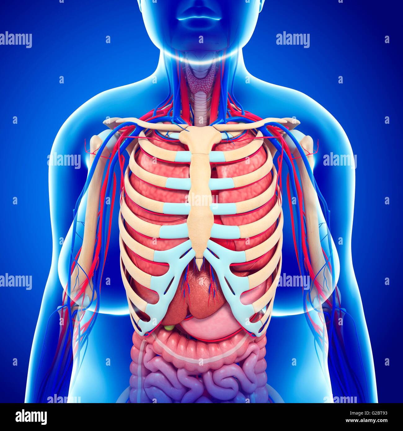
The rounded ends are attached at joints to the thoracic vertebrae posteriorly and the flattened ends come together at the sternum anteriorly. With each succeeding rib, from the first, or uppermost, the curvature of the rib cage becomes more open. Of all human pancreas anatomy. Others attach indirectly because they are attached to the cartilage of the rib above. The lungs are two separate but connected organs located in the upper chest, covered by the rib cage.
Human Ribcage And Internal Organs Illustration Stock Photo Alamy from c8.alamy.com Related posts of rib cage diagram with organs abdominal cavity chart. The human rib cage is made up of 12 pairs of ribs, some of which attach to a bony process in the front of the chest called the sternum. The thoracic cage protects the heart and lungs. 3d illustration showing organs of digestive system with highlighted pancreatic gland, view from back The ribs form the main structure of the thoracic cage protecting the thoracic organs, however their main function is to aid respiration3. It functions as protection for the vital organs of the chest, such as the heart and lungs. Inner body cardiovascular system 7 photos of the inner body cardiovascular system digestive system cardiovascular system, human anatomy cardiovascular system, human body cardiovascular system, human body circulatory system, human body lymphatic system, human body nervous system, human body respiratory system, inner. Two of the most notable organs behind the left side of the rib cage are the left lung and the spleen.
The ribs partially enclose and protect the chest cavity, where many vital organs (including the heart and the lungs) are located.
The thoracic cage (rib cage) is the skeletal framework of the thoracic wall, which encloses the thoracic cavity. It functions as protection for the vital organs of the chest, such as the heart and lungs. The thoracic cage, also called the rib cage. Two of the most notable organs behind the left side of the rib cage are the left lung and the spleen. The right upper quadrant of the abdomen includes the pancreas, right kidney, gallbladder, liver, and intestines. The thoracic cage surrounds and protects the heart and lungs in the thoracic cavity. The human rib cage is made up of 12 paired rib bones; Surface anatomy of abdominal organs and ribcage of the human body in this image, you will find jugular notch, aorta, inferior vena cava, the upper border of the l1 celiac trunk, transpyloric plane, lower border of the l1 superior mesenteric artery, an l2 approximate origin of a renal artery in it. Heart in thorax, highlighting the valves. Each are symmetrically paired on a right and left side. Inner body cardiovascular system 7 photos of the inner body cardiovascular system digestive system cardiovascular system, human anatomy cardiovascular system, human body cardiovascular system, human body circulatory system, human body lymphatic system, human body nervous system, human body respiratory system, inner. The rib cage is collectively made up of long, curved individual. 3d illustration showing organs of digestive system with highlighted pancreatic gland, view from back
Although that is one key function, the ribcage does so much more. The lungs are two separate but connected organs located in the upper chest, covered by the rib cage. Gross anatomy there are 12 pairs of ribs which are separated by intercostal spaces. The transpyloric plane and mcburney's point are among the marked locations. Rib cage anatomy skeleton anatomy human muscle anatomy human anatomy anatomy organs anatomy and physiology xiphoid process avascular necrosis forensics.
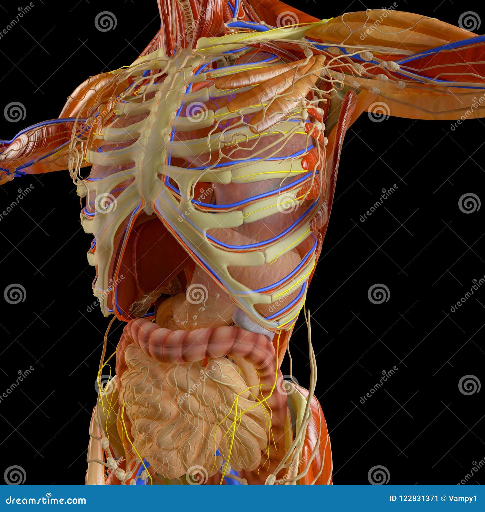
Human Body X Ray View Of The Respiratory Apparatus And Digestive Tract In The Ribcage Anatomy Stock Illustration Illustration Of Digestive Contraction 122831371 from thumbs.dreamstime.com The thoracic cage surrounds and protects the heart and lungs in the thoracic cavity. Inner body cardiovascular system 7 photos of the inner body cardiovascular system digestive system cardiovascular system, human anatomy cardiovascular system, human body cardiovascular system, human body circulatory system, human body lymphatic system, human body nervous system, human body respiratory system, inner. The ribs partially enclose and protect the chest cavity, where many vital organs (including the heart and the lungs) are located. Surface projections of the major organs of the trunk, using the vertebral column and rib cage as main reference points of superficial anatomy. The first seven ribs p. Of all human pancreas anatomy. There is one last component of the axial skeleton we did not cover last lab: The top rib bones are thin, curved bones that connect to the sternum.
The primary responsibilities of the ribcage involve protecting the thoracic visceral organs, enclosing the thoracic visceral organs, and is included in the general mechanics of the process of breathing.
The human rib cage is made up of 12 pairs of ribs, some of which attach to a bony process in the front of the chest called the sternum. Related posts of rib cage organs anatomy inner body cardiovascular system. Middle back and rib pain can also be caused by organs under your ribs like kidney stones, gallbladder pain, or lung disease. Surface projections of the major organs of the trunk, using the vertebral column and rib cage as main reference points of superficial anatomy. Related posts of rib cage diagram with organs abdominal cavity chart. Of all human pancreas anatomy. There is one last component of the axial skeleton we did not cover last lab: The transpyloric plane and mcburney's point are among the marked locations. Format_list_bulleted contents add the ribs are a set of twelve paired bones which form the protective 'cage' of the thorax. Both the liver and the stomach are located in the lower chest region under the thoracic diaphragm, a sheet of muscle at the bottom of the rib cage that separates the chest cavity from the abdominal. The rib cage is collectively made up of long, curved individual. Heart in thorax, highlighting the valves. The thoracic cage (rib cage) is the skeletal framework of the thoracic wall, which encloses the thoracic cavity.
Related posts of rib cage organs anatomy inner body cardiovascular system. With each succeeding rib, from the first, or uppermost, the curvature of the rib cage becomes more open. The first seven ribs attach directly to the sternum through cartilage that forms at the end of each rib. In this video we discuss the structure of the rib cage or thoracic cage. Surface anatomy of abdominal organs and ribcage of the human body in this image, you will find jugular notch, aorta, inferior vena cava, the upper border of the l1 celiac trunk, transpyloric plane, lower border of the l1 superior mesenteric artery, an l2 approximate origin of a renal artery in it.
What Are Some Characteristics Of The Organs Under The Left Side Of The Rib Cage Quora from qph.fs.quoracdn.net The first seven ribs p. The rounded ends are attached at joints to the thoracic vertebrae posteriorly and the flattened ends come together at the sternum anteriorly. The human rib cage is made up of 12 paired rib bones; The thoracic cage (rib cage) forms the thorax (chest) portion of the body. Abdominal cavity chart 14 photos of the abdominal cavity chart abdominal cavity cancer, abdominal cavity contains, abdominal cavity diagram picture, abdominal cavity pain, abdominal cavity quadrants, abdominal cavity regions, air in abdominal cavity, fluid buildup in abdominal cavity, stomach, abdominal cavity cancer. The right upper quadrant of the abdomen includes the pancreas, right kidney, gallbladder, liver, and intestines. Heart in thorax, highlighting the valves. Others attach indirectly because they are attached to the cartilage of the rib above.
The lungs are two separate but connected organs located in the upper chest, covered by the rib cage.
We cover the different bones that make up the rib cage and some of the functions of. The right upper quadrant of the abdomen includes the pancreas, right kidney, gallbladder, liver, and intestines. The top rib bones are thin, curved bones that connect to the sternum. It consists of 24 rib bones, along with the sternum, 12 thoracic vertebrae and costal cartilages. The ribs form the main structure of the thoracic cage protecting the thoracic organs, however their main function is to aid respiration3. It consists of the 12 pairs of ribs with their costal cartilages and the sternum (). With each succeeding rib, from the first, or uppermost, the curvature of the rib cage becomes more open. As part of the bony thorax, the ribs protect the internal thoracic organs. 3d illustration showing organs of digestive system with highlighted pancreatic gland, view from back The first seven ribs p. The thoracic cage protects the heart and lungs. Gross anatomy there are 12 pairs of ribs which are separated by intercostal spaces. The human rib cage is made up of 12 pairs of ribs, some of which attach to a bony process in the front of the chest called the sternum.
Of all human pancreas anatomy rib cage anatomy. We cover the different bones that make up the rib cage and some of the functions of.